Ultrasound Unit
Ultrasound Unit
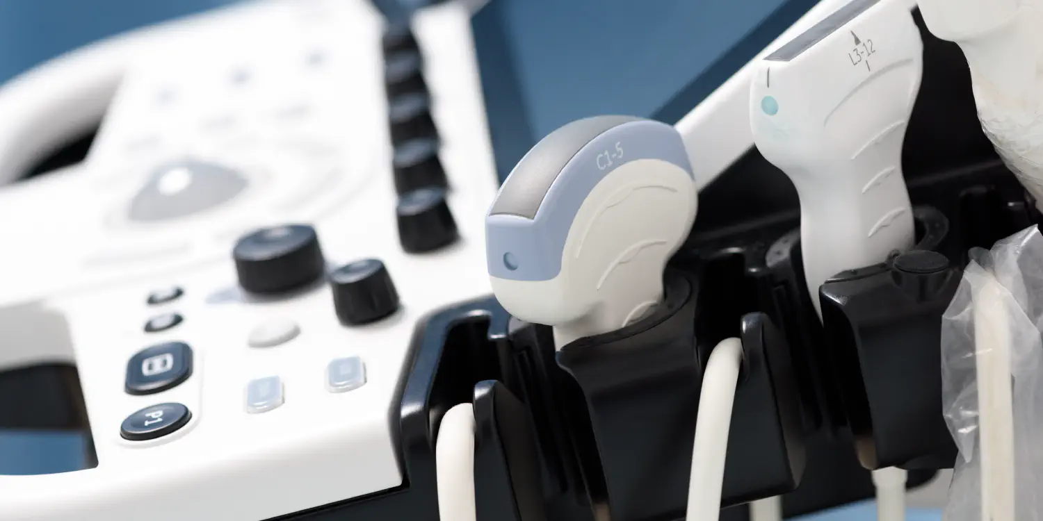
Ultrasound is often the first diagnostic tool a clinician reaches for. It’s fast, radiation-free, and gives real-time images of soft tissue, organs, blood vessels, and the heart that drive clinical decisions across almost every specialty. At Oasis Clinics, all ultrasound scans are performed by physicians with specialized training in ultrasound diagnostics, using the latest imaging equipment.
Because the ultrasound unit is housed within the same building as the cardiology, gastroenterology, OB/GYN, endocrinology, and vascular teams, scans can frequently be done on the same day as your consultation, with findings feeding directly into the clinical discussion.
General Diagnostic Scans
Abdominal Ultrasound
Visualizes the liver, gallbladder, kidneys, spleen, pancreas, and abdominal aorta. Commonly ordered for:
- Abdominal or flank pain
- Suspected gallstones or gallbladder disease
- Elevated liver enzymes on blood tests
- Kidney abnormalities found incidentally or on lab work
- Screening for abdominal aortic aneurysm in at-risk patients
Pelvic Ultrasound
Examines the uterus, ovaries, and bladder in women, and the bladder and prostate in men. Used for gynecological assessment, fertility investigation, and urological evaluation.
Thyroid Ultrasound
Assesses the thyroid gland for nodules, cysts, goiter, and structural abnormalities. Typically performed alongside thyroid blood tests (TSH, T3, T4) when thyroid dysfunction is suspected or already confirmed. Thyroid disease is among the most prevalent endocrine conditions in Egypt and the wider region, and ultrasound is the definitive structural assessment.
Breast Ultrasound
Detailed imaging of breast tissue for:
- Evaluating a palpable lump or area of concern
- Characterizing a mass identified on mammography
- Assessment in younger patients where high breast density makes mammography less informative
Cardiovascular and Vascular Scans
Echocardiogram
A cardiac ultrasound that produces real-time images of the heart’s chambers, valves, and wall motion. Used to assess heart function, detect valve abnormalities, measure ejection fraction, and monitor the heart’s response to treatment. Performed in coordination with the Oasis Clinics cardiology team.
Carotid Ultrasound
Examines the carotid arteries in the neck for plaque buildup, narrowing, and blood flow abnormalities. A key tool in stroke risk assessment, ordered for patients with cardiovascular risk factors, a history of TIA (transient ischemic attack), or as part of a preventive cardiac workup.
Doppler Ultrasound
Measures and visualizes blood flow through arteries, veins, and organs in real time. Used to detect vascular blockages, assess organ perfusion (liver, kidneys), and evaluate graft or transplant blood flow.
Venous Ultrasound (Extremities)
Examines the veins of the arms and legs for deep vein thrombosis (DVT), venous insufficiency, and the structural source of varicose veins. Typically arranged by the vascular team when leg swelling, pain, heaviness, or visible varicosities need investigation.
Specialized Ultrasound
Musculoskeletal Ultrasound
Real-time imaging of tendons, ligaments, muscles, and joints. Used to diagnose tendon tears, muscle injuries, joint effusions, and bursitis. Also used to guide injection therapy into painful joints and soft tissue structures with greater accuracy than landmark-based technique. Particularly useful alongside physiotherapy assessment for sports injuries and overuse conditions.
Ultrasound-Guided Needle Placement
Ultrasound guidance allows a needle to reach a specific target with precision, in real time, reducing procedural risk and improving accuracy. Used for:
- Biopsies of thyroid nodules, breast masses, and abdominal lesions
- Drainage of fluid collections
- Joint and bursa injections
One Visit, Not Three
Most ultrasound scans at Oasis Clinics can be arranged the same day as a specialist consultation. You don’t need to book a separate appointment at a different facility, wait for a report to arrive by fax, and then return to your doctor to discuss it. The scan, the specialist interpretation, and the clinical conversation happen in sequence, on the same visit. For patients managing a condition across multiple specialties, that efficiency is built into how the unit operates.